
Sample Long-Term Potentiation and Depression Research Paper. Browse other research paper examples and check the list of research paper topics for more inspiration. If you need a research paper written according to all the academic standards, you can always turn to our experienced writers for help. This is how your paper can get an A! Feel free to contact our research paper writing service for professional assistance. We offer high-quality assignments for reasonable rates.
Long-term potentiation (LTP) is a long-lasting increase in postsynaptic response seen at glutamate synapses following afferent activation. In the laboratory, LTP typically is induced by a brief tetanus to afferents and results in a larger postsynaptic response to a constant input that lasts for an hour or more. Long-term depression (LTD) is a long-lasting decrease in the postsynaptic response seen at the same synapses. LTD is induced by a low-frequency tetanus leading to a postsynaptic response depression lasting for an hour or more. Both LTP and LTD are alterations in synaptic gain that display an induction threshold, ar e specific to the synapses activated, are induced by Ca²⁺ entering the postsynaptic cell, and display associativity—such that the pairing of a strong and weak (unable to induce change alone) stimulus will induce gain change in both inputs. Both forms of synaptic plasticity are thought to be central to the formation of neural networks underlying information storage. The commonly accepted model of the storage of experiential information (e.g., memory) involves the establishment of networks of neurons interconnected in such a way to encode the salient aspects of the experience. These networks—which may be quite extensive and involve thousands of neurons—are themselves the neural representation of experience. LTP and LTD are found in brain regions and at developmental times that suggest that they may encode far more than ongoing experience, perhaps being generic synaptic mechanisms involved in the establishment of fundamental cortical sensory and motor representation, among other things. Cortical LTP is most pronounced during early development coinciding with the critical period for environmentallyinduced synaptic plasticity, suggesting that the same mechanism may underlie both phenomena.
Academic Writing, Editing, Proofreading, And Problem Solving Services
Get 10% OFF with 24START discount code
1. Cortical Distribution
LTP/LTD are found throughout the neocortex at glutamatergic synapses of pyramidal cells and interneurons. One or both of these forms of synaptic plasticity have been reported in primary sensory areas, primary motor areas, and ‘association’ cortex of rodent, cat, and primate brain, as well as cortical projections to subcortical structures (thalamus, striatum). While there are some differences between cortical areas, generally both LTP and LTD are seen at all locations, as in hippocampus (but unlike cerebellar Pyrkinje cell synapses where LTD predominates). As opposed to the hippocampus, where most studies of synaptic plasticity are performed, the complex anatomy of cortex makes it difficult to stimulate monosynaptic, homogeneous afferent pathways. Most studies, either in itro or in i o, tetanize the white matter afferents and record the postsynaptic effects in corresponding cortical column, or stimulate and record intracortically within the column (layer IV/V to II/ III or layer II to V). In both cases, stimulation activates a heterogeneous population of afferents and antidromically activates efferents traversing the region, making interpretation of the synapses involved problematic. Similarly, the postsynaptic responses are often combinations of orthodromic monoand polysynaptic responses and antidromic responses.
2. Mechanisms of nmdaLTP
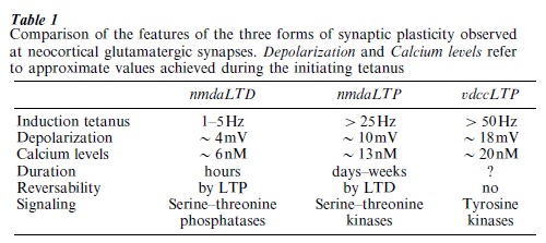
There are two different forms of cortical LTP: one mediated by activation of the NMDA glutamate receptor subtype, the other mediated by activation of Voltage Dependent Calcium Channels (VDCCs). Both cortical forms appear to have the same characteristics as LTP found in hippocampus, namely: glutamate receptor subtype or VDCC involvement, input sp ecificity, associativity, activation threshold and Ca²⁺ dependency. The induction of the form of LTP mediated by the NMDA receptor (nmdaLTP) requires afferent activity associated with postsynaptic depolarization via the AMPA receptor (a ligand-gated glutamate receptor gating Na⁺/ K⁺) sufficient to activate the ligand and voltage sensitive NMDA receptor (NMDAR). The active NMDAR gates Ca into the postsynaptic cell where it activates several serine– threonine kinases (PKA, PKC, CaMKII) leading to the phosphorylation of the AMPAR—resulting in larger EPSPs, and the activation of incompletely understood signal transduction cascades (involving the CREB and Ras-MAPK cascades) ultimately leading to an alteration in gene expression protein synthesis. Most of the studies investigating the signaling pathways in LTP have been done in hippocampal neurons using both enzyme inhibitors, and developmental or targeted inducible genetic knock-out animals. The relatively few neocortical studies indicate that the same mechanisms are present. The activated kinases, some of which are autophosphorylated as a result of their activation (thus ensuring their continued activity in the absence of the calcium transient), serve to phosphorylate their substrates. Many of the receptors, channels, and enzymes present postsynaptically have phosphorylation sites which, when activated, result in increases in postsynaptic responsivity. The expression of LTP immediately following a tetanus is primarily due to phosphorylation of the AMPAR. As will be seen later, some phosphorylation sites on the AMPAR and other substrates are reversible and can be dephosphorylated through the action of phosphatases associated with LTD. It is thought that the persistence of LTP involves synthesis of synaptic proteins, as enzyme inhibitors and protein synthesis inhibitors limit the duration of LTP expression (in hippocampus).
3. Mechanisms of vdccLTP
The form of LTP mediated by VDCCs (vdccLTP) is initiated by depolarizing the postsynaptic cell beyond that required for nmdaLTP—to activate L-type VDCCs and allow an influx of Ca²⁺ into the postsynaptic cell. This means that either the degree of afferent activity must lead to a higher level of postsynaptic depolarization or that other elements must assist in the establishment of appropriate levels of depolarization to induce this form of LTP. There are two known means by which cellular processes contribute to the induction of vdccLTP. The first is through the simultaneous induction of nmdaLTP which contributes depolarization to the postsynaptic cell and thus helps to achieve the conditions for the activation of VDCCs. The second is by reducing the inhibitory drive onto the postsynaptic cell to facilitate the induction vdccLTP. As compared to hippocampus, neocortical LTP appears to be regulated to a greater extent by enhanced inhibitory tone, as GABAa blockers are often required to elicit either form of cortical LTP in itro. The cellular targets of Ca²⁺ gat ed through VDCCs appears to be different from Ca²⁺ gated through NMDAR channels, in that enzyme inhibitors of serine threonine kinases that block nmdaLTP have no affect on the induction or expression of vdccLTP. vdccLTP depends upon the activation of tyrosine kinases, and involves the participation of tyrosine receptor kinases (trk)—the receptors for neurotrophins. In visual cortex of young rats, the application of the neurotrophin BDNF facilitates the induction of synaptic plasticity to a weak tetanus. As opposed to nmdaLTP which can be reversed (depotentiated) by LTD induction, vdccLTP appears to be irreversible. Since both forms of LTP are induced by afferent activity leading to postsynaptic depolarization, sufficient activity will lead to the expression of both forms simultaneously.
The two forms of LTP have somewhat different cortical distribution, with nmdaLTP found at intracortical synapses and associated with white matter activation early in development. In adult cortex, NMDARs decline in number and nmdaLTP seen to white matter stimulation is reduced or absent, leaving vdccLTP dominant. A similar, but less pronounced, effect is seen in hippocampus as well.
4. Long-term Depression
LTD is the decremental counterpar of LTP whose mechanism of action also utilizes Ca²⁺ gated through NMDARs. The evidence from Ca imaging studies suggests that the amount of Ca entering the postsynaptic cell is critical in determining which form of synaptic plasticity will be expressed. LTP induction seen following high frequency afferent activity is associated with greater postsynaptic cytosolic Ca²⁺ levels than that seen following prolonged low frequency activation leading to LTD. These low levels of calc ium in the postsynaptic cell bind to high-affinity Ca²⁺ binding sites on protein phosphotases, whose action is to dephosphorylate the substrates of serine threonine kinases. Thus, LTD acts to dephosphorylate sites that have been phosphorylated by the action of protein kinases (the removal of nmdaLTP by LTD induction is termed depotentiation). Thus, nmdaLTP and LTD (or phosphorylation and dephosphorylation) exist in a opposing regulatory relationship.
5. Properties of Plastic Cortical Synapses
Studies in hippocampus have revealed several other forms of synaptic plasticity that have not been reported for neocortex. These include a presynaptic form of LTP at hippocampal CA3 synapses and a form of LTP utilizing opiate transmitters. Given the relative paucity of mechanistic studies of LTP in neocortex, it is unclear if these forms are absent or undiscovered. Two additional aspects of LTP seen in hippocampus are present in at least some regions of neocortex: the influence of the metabotropic glutamate receptor (mGluR) and the presence of the retrograde messenger NO. The mGluRs are a family of G-protein linked glutamate receptors whose different effects are mediat ed by PKC activation and IP3-mediated release of Ca²⁺ from postsynaptic endoplasmic reticuluum stores. Various mGluR antagonists and knock-outs have demonstrated the requirement for mGlu involvement in LTP in both hippocampus and neocortex, although it’s precise role remains elusive. There may be regional cortical differences however, as some mGluR antagonists are ineffective in blocking PI hydrolysis and synaptic plasticity in visual cortex. As in hippocampal pyramidal cell synapses, there is evidence for the involvement of the retrograde messenger NO in neocortical synapses, as inactivation of its synthetic enzyme blocks LTP in auditory and somatosensory cortex.
As changes in synaptic gain are initiated by incremental increases in postsynaptic depolarization (with increasing depolarization, nmdaLTD, then nmdaLTP, then vdccLTP are induced), modulatory synaptic influences onto cortical pyramidal cells can be expected to influence the form and degree of synaptic plasticity induced by glutamatergic activity. Studies primarily in visual cortex have demonstrated: the inhibiting effect of mACh antagonists on LTP, the inhibiting effect of 5-HT1A agonists on visual cortex LTP, and the dopamine facilitation of intracortical prefrontal cortex LTD. As different cortical regions have differential neuromodulatory inputs, it might be expected that they will influence synaptic plasticity in different directions. More detailed studies in hippocampus have revealed that no two synapse types share identical properties. While they are outwardly similar, they differ mechanistically. The same may be found across neocortical synapses.
In sum, neocortical synaptic plasticity is pheno- menologically and mechanistically similar to that seen in the better studied archicortex (hippocampal formation). Differences are seen primarily in the regional distribution of forms of LTP between cortical areas and within cortical laminae and the relative dominance of the VDCC form of LTP, particularly its prevalence with maturity. LTP LTD remain the best candidates for processes that alter the synaptic gain at nodes in neural networks encoding information—and thus defining the architecture of the network. If the forms of synaptic plasticity seen in neocortex underlie information storage, it must be demonstrated that they are specific and necessary to the process they govern. Beyond this, consideration must be given to the nature of the information that is stored, how it is reaccessed, and how it integrates with other brain functions.
Bibliography:
- Aroniadou V A, Keller A 1995 Mechanisms of LTP induction in rat motor cortex in vitro. Cerebral Cortex 5: 353–62
- Bear M F 1996 A synaptic basis for memory storage in the cerebral cortex. Proceedings of the National Academy of Science USA 93: 13453–9
- Bear M F, Kirkwood A 1993 Neocortical long-term potentiation. Current Opinion Neurobiology 3: 197–202
- Kirkwood A, Dudek S M, Gold J T, Aizenman C D, Bear M F 1993 Common forms of synaptic plasticity in the hippocampus and neocortex in vitro. Science 260: 1518–21
- Lisman J E, Fallon J R 1999 What maintains memories? Science 283: 339–40
- Singer W 1995 Development and plasticity of cortical processing architectures Science 270: 758–64
ORDER HIGH QUALITY CUSTOM PAPER

Always on-time

Plagiarism-Free

100% Confidentiality